Article Text

Abstract
Introduction Intra-articular injections of mesenchymal stromal cells concentrates showed promising results in the treatment of knee osteoarthritis (OA). Among these, bone marrow aspirate concentrate (BMAC) has been widely adopted in clinical practice. More recently, microfragmented adipose tissue (MFAT) has been proposed as a more suitable solution. However, there is still no high-level evidence demonstrating the superiority of MFAT to BMAC. The aim of this randomised controlled trial is to compare the safety and clinical outcomes of a single intra-articular injection of BMAC versus a single intra-articular injection of MFAT in patients with knee OA.
Methods and analysis 204 patients aged 40–75 years and affected by knee OA are randomised to receive a single injection of BMAC or MFAT in a 1:1 ratio. The primary outcome of the study is the Western Ontario and McMaster University OA index (WOMAC) pain score at 6 months. The secondary outcomes of the study are the WOMAC pain score at 2 months and 12 months and the WOMAC subscales, the total WOMAC score, the International Knee Documentation Committee subjective and objective scores, the Knee Injury and OA Outcome score, the visual analogue scale (VAS) for pain evaluation, the EuroQol VAS and the Tegner score at 2 months, 6 months and 12 months. Moreover, the study aims at demonstrating whether these products have disease-modifying effects: radiographs and magnetic resonance evaluations are performed at baseline and at 12 months of follow-up, while systemic OA biomarkers are evaluated at baseline and after 2 months, 6 months and 12 months. As a tertiary outcome, this study aims at identifying the factors that influence the clinical response, including baseline patient clinical characteristics, biological features of the OA joint, as well as anabolic and anti-inflammatory properties of the injected products.
Ethics and dissemination The study protocol has been approved by Emilia Romagna’s Ethics Committee Comitato Etico Area Vasta Emilia Centro (CE-AVEC), Bologna, Italy (protocol number: 150/2023/Sper/IOR). Written informed consent is obtained from all participants. The findings of this study will be disseminated through peer-reviewed publications and conference presentations.
Protocol version March 2023.
Trial registration number NCT06040957.
- Orthopedics
- Knee
- Randomized Controlled Trial
- Mesenchymal Stem Cells
- Patient Reported Outcome Measures
This is an open access article distributed in accordance with the Creative Commons Attribution Non Commercial (CC BY-NC 4.0) license, which permits others to distribute, remix, adapt, build upon this work non-commercially, and license their derivative works on different terms, provided the original work is properly cited, appropriate credit is given, any changes made indicated, and the use is non-commercial. See: http://creativecommons.org/licenses/by-nc/4.0/.
Statistics from Altmetric.com
STRENGTHS AND LIMITATIONS OF THIS STUDY
This is a prospective, randomised controlled trial (RCT) performed in a highly specialised orthopaedic centre for cartilage preservation procedures and knee osteoarthritis (OA) treatment.
Patients are analysed using patient-reported outcome measures, objective measures, X-rays, MRI and biomarkers evaluation.
The analysis of patient baseline characteristics and disease-related factors can help better define the aspects that make different individuals more or less responsive to these treatments.
The main limitation of this RCT is the inability to maintain patients blinded to the treatment allocation due to the different incisions performed for each treatment group.
The 12 months of follow-up timeframe may not be sufficient to analyse the clinical and biological response to the two treatments and the progression of knee OA over time.
Introduction
Knee osteoarthritis (OA) is an increasingly common condition causing joint pain and functional limitation, often requiring invasive surgical procedures like knee replacement. These have long rehabilitation and potential severe morbidity; they often need reoperation, and overall, they present extensive socioeconomic impact.1 2 The only partially satisfactory results of these treatments led to the development of less invasive procedures such as intra-articular injections of orthobiologics. Among these, mesenchymal stromal cells (MSCs) are emerging as a promising solution, given their biologic potential, reported both for cultured cells and minimally manipulated products, with preclinical studies documenting both anti-inflammatory and anabolic properties of MSCs.3–5 Aiming at reducing costs and avoiding regulatory restrictions associated with cell culture approaches, minimally manipulated products have become a popular strategy to exploit the potential of MSCs concentrates directly on-site in a one-step treatment, while avoiding isolation and cell culture before the injection.6 7 Among the main treatment modalities that emerged in the past few years, bone marrow aspirate concentrate (BMAC) has been proposed as an injective approach for degenerative orthopaedic conditions like knee OA.8 However, despite improvements in clinical and functional scores, overall results are still suboptimal, and preclinical studies supporting the rationale of BMAC injections are scarce, with an overall low quality of evidence of the clinical studies.8 More recently, minimally manipulated adipose tissue (MM-AT) derived products and, in particular, microfragmented adipose tissue (MFAT), have been proposed as a more promising alternative for the treatment of OA, given the possible advantages of adipose tissue over other sources of MSCs isolated from different tissues such as bone marrow and synovial tissue.9–13 In fact, MM-AT derived products represent a valid source of MSCs given the tissue abundance, ease of harvesting with little patient discomfort, high concentration in MSCs, but also their ability to respond to an inflammatory environment better than BMAC in preclinical studies.14 However, as of today, no randomised controlled trial (RCT) directly compared the clinical benefit of BMAC and MFAT orthobiologic approaches, and the current literature provides poor guidance being limited by low level studies, heterogeneity in treatment protocols and overall poor reporting of treatment characteristics and results in terms of both subjective and objective outcomes.
The hypothesis is that MFAT injections can provide better clinical outcomes compared with BMAC injections for the treatment of patients with symptomatic knee OA. This project will provide, for the first time, high-level evidence to confirm this hypothesis in the clinical setting.
Objectives and trial design
A RCT was designed to compare efficacy and safety of a single intra-articular injection of BMAC versus a single intra-articular injection of MFAT to address patients with knee OA, with a 1:1 allocation ratio. As secondary goals, this study aims at demonstrating whether BMAC or MFAT injections can have disease-modifying effects by investigating tissue modifications through combined imaging and biological evaluations. As a tertiary goal, this study aims at identifying the factors that influence the clinical response, including baseline patient clinical characteristics and biological features of the OA joint, as well as anabolic and anti-inflammatory properties of the injected products.
Methods and analysis
Study setting
The study is a single-centre RCT, with all activities related to the study performed at a single site (IRCCS Istituto Ortopedico Rizzoli, Bologna, Italy). This trial protocol is produced according to the Standard Protocol Items: Recommendations for Interventional Trials (SPIRIT) reporting guidelines.15
Patient and public involvement
Patients were not involved in planning research questions, outcome measures or design of the study.
Eligibility criteria
Patients are recruited according to the following criteria.
Inclusion criteria
Men or women aged between 40 years and 75 years, to reduce heterogeneity and in light of the possible lower potential of cells from older patients.16
Symptomatic tibiofemoral OA with a history of knee pain and/or swelling for at least 6 months.
Radiographic signs of OA (grade 1–4 according to the Kellgren-Lawrence classification).
Ability and consent to participate in clinical and imaging follow-ups.
Informed consent signed.
Exclusion criteria
Patients with cognitive impairment or unable to provide informed consent.
Patients with active malignancy.
Patients suffering from rheumatic diseases or arthritis secondary to other inflammatory diseases.
HIV infection or viral hepatitis.
Patients suffering from uncontrolled diabetes or thyroid disorders.
Patients with a history of alcohol or drug abuse.
Pregnancy, breast-feeding or intention to start pregnancy during the study.
Patients who underwent knee surgery in the previous 12 months.
Lower limb axial deviation >5° on the frontal plane, evaluated on a full-length standing radiograph.
History of major knee trauma within 6 months prior to the treatment.
History of intra-articular injections within 6 months prior to the treatment.
Patients with a history of allergic reactions to local anaesthetics used in the procedures.
Presence of joint infection or other knee lesions causing pain other than OA (eg, osteochondral lesions, impairing meniscal tears causing mechanical symptoms).
Body mass index (BMI) >35 kg/m2, to avoid the possible proinflammatory state related to the adipose tissue from more obese patients.14
Intervention
Patients are treated by orthopaedic surgeons with experience in orthobiologics. The procedure is performed in a single step in the operating room with patients in supine position, with a sedative and analgesic oral premedication of tramadol 100 mg/mL and bromazepam 2.5 mg/mL adjusted for weight (number of oral drops is equal to weight in kg divided by 3) and using local anaesthesia.
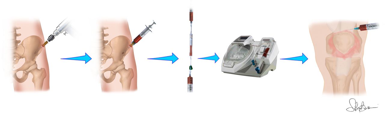
In the BMAC group, bone marrow is harvested from the anterior iliac crest: a single incision is made at the level of the anterior iliac crest using a dedicated European Community (EC) approved kit (Isto Biologics Magellan) and collected using two 30 mL syringes coated with heparin for a total of 60 mL. This anatomical site has been proven to be one of the most appropriate in terms of biological potential.16 The harvested bone marrow is filtered with a heparin washed filter and then centrifuged through the Magellan centrifuge (Isto Biologics, Hopkinton, Massachusetts, USA, previously Arteriocyte Medical Systems, Hopkinton, Massachusetts, USA) at a rate of 3600 RPM for 15 min, thus obtaining 8 mL of BMAC (figure 1). The incision is then sutured using a single stitch, which has to be removed 2 weeks after the surgery.

Bone marrow aspirate concentrate injection procedure.
In the MFAT group, adipose tissue is harvested from the subcutaneous abdominal fat, as this site proved to be the most appropriate in terms of biological potential and ease of harvesting.9 After local anaesthesia, a subcentrimetric incision is performed on both sides of the lower or lateral abdomen. Before harvesting the fat, each side is injected with 180 mL of Klein solution (1 mL of 2 μg/ml epinephrine and 40 mL of 0.02% lidocaine in 500 mL of saline solution) using a disposable 17-gauge blunt cannula connected to a 60-millilitre Luer Lock syringe. Adipose tissue is then collected using a 13-gauge blunt cannula, for fast and atraumatic suction, connected to a 20-millilitre Vac Lok syringe. The harvested fat is immediately processed using the Lipogems system (Lipogems International Spa, Milan, Italy) as previously described.17 The entire process is performed in complete immersion in isotonic solution, thus minimising cell trauma. The size of adipose tissue clusters is progressively reduced with a mild mechanical action to microspheres, in accordance with the manufacturer’s instructions. This process allows for the elimination of oily substances, cell debris and blood residues. Finally, the resulting microfragmented tissue (8 mL) is collected in a 10-millilitre syringe.18 The process is shown in figure 2. The two incisions are then sutured using a single stitch for each side, which has to be removed 2 weeks after the surgery. After the surgery, a girdle must be worn for 10 days.

Microfragmented adipose tissue injection procedure.
An arthrocentesis is then performed in the affected knee to collect synovial fluid (SF). This will not be possible in all patients as SF may be absent or insufficient. In addition, if synovitis is present, a synovial membrane biopsy is collected with ultrasound guidance with a dedicated biopsy needle. In both groups, the intra-articular injection of 6 mL of orthobiologic products is then performed with ultrasound guidance in a lateral suprapatellar approach using an 18-gauge needle, with the patient in supine position and the knee fully extended. The remaining 2-millilitre sample is sent to the laboratory for the in vitro analyses together with the SF and synovial tissue samples.
At the end of the injection, the patient is encouraged to bend and extend the knee a few times to allow the product to spread throughout the joint. The postoperative protocol includes rest and avoiding high-impact sports activities and strenuous work for 2 weeks, without restrictions in weight-bearing following the procedure. After the removal of stitches, gradual return to sport is allowed as tolerated, with exercise bike and aquatic therapy being the recommended activities.
Outcomes
Patients are evaluated at baseline and at 2 months, 6 months and 12 months with validated questionnaires. The primary outcome of the study is the Western Ontario and McMaster University Osteoarthritis index (WOMAC) pain score at 6 months. The secondary outcomes of the study are the WOMAC pain score at 2 months and 12 months, the WOMAC subscales (function, stiffness), the total WOMAC score, the International Knee Documentation Committee subjective and objective scores, the Knee Injury and Osteoarthritis Outcome score, the visual analogue scale (VAS) for pain evaluation, the EuroQol VAS for the overall quality of life evaluation and the Tegner score to document the activity level of the treated patients at 2 months, 6 months and 12 months.
Imaging evaluation is performed by analysing knees with radiographs (anteroposterior and lateral views) at baseline and at the 12-month follow-up to assess the OA grade according to the Kellgren-Lawrence classification. An approved artificial intelligence imaging analysis system is also used to determine the Kellgren-Lawrence grade. High-resolution 3.0 Tesla MRI is performed at baseline and at the 12-month follow-up, and the Whole-Organ MRI Score is used to assess articular cartilage morphology, bone marrow oedema, subchondral cysts, articular profile, marginal osteophytes, meniscal integrity and synovitis.
Systemic OA biomarkers are evaluated at baseline and after 2 months, 6 months and 12 months of follow-up, through circulating microRNAs (miRNAs) expression analysis and spontaneous osteoclastogenesis evaluation. Circulating miRNAs from peripheral blood samples are analysed as reported below. An aliquot of peripheral blood sample (approximately 5 mL) is stored at 4°C overnight, then centrifuged and plasma is then stored at −80°C. Circulating miRNAs are isolated following the guidelines of the commercial kit of extraction and isolation (mirVana miRNA Isolation Kit), which can provide highly sensitive results by enabling the miRNA detection from just 1 pg starting material. MiRNA characterisation is performed after retrotranscription of miRNAs isolated using the TaqMan Advanced miRNA cDNA Synthesis Kit. MiRNAs involved in OA progression or inflammation are identified and quantified using precast miRNA seq panel analysis (TaqMan Advanced miRNA Human A and B 96-well Plates, fast).19–23 Data obtained are analysed with bioinformatic and multivariate analyses. Subsequently, a list of putative biomarkers is identified by means of bioinformatic investigations.
Spontaneous osteoclastogenesis is evaluated. Monocytes from patients with OA display enhanced capacity to generate osteoclasts (OCs) compared with cells from healthy controls.24 OCs are obtained from approximately 2 mL of peripheral blood from each patient. More precisely, peripheral blood mononuclear cells (PBMCs) are isolated with Ficoll density-gradient centrifugation and then cultured in alpha-minimum essential medium. After monitoring viability, once a week for 3 weeks using Alamar blue dye test, a differentiation assay is performed after 21 days of culture through tartrate-resistant acid phosphatase staining; the large, multinucleated cells (>3 nuclei), which developed a brown colour, are scored as positive cells, and the ratio between the brown-coloured region and total image area is measured using an image analysis system of inverted microscope. In addition, the supernatants of cells, after 21 days of culture, are stored at −80°C and evaluated for cathepsin K, metalloproteinase-7 (MMP-7) and MMP-9 production, with immunoenzymatic ELISA tests (minimum detectable dose less than 0.057 ng/mL), for OC activity assessment. Finally, the resorption assay is carried out by culturing PBMCs on bone slices, then stained with toluidine blue staining to reveal pits, and an image analysis programme evaluates the resorption area.
At baseline, autologous BMAC and MFAT remaining from patient treatment (approximately 2 mL) are collected and transferred aseptically to the laboratory for in vitro analysis. An aliquot is immediately cultured for the evaluation of cell viability and protein production. The Alamar blue dye test is used for cell viability assessment and the production of the most important factors involved in trophic and anti-inflammatory processes and interleukins (ILs), such as platelet-derived growth factor, transforming growth factor β, vascular endothelial growth factor (VEGF), fibroblast growth factor, insulin-like growth factor-I, granulocyte-macrophage colony-stimulating factor, bone morphogenetic protein 2 (BMP2), BMP7, IL1ß, IL6, IL8 and IL-1ra, evaluated from the supernatant using Bio-Plex Pro panels.
The remaining aliquot of BMAC and MFAT is employed for MSC characterisation. After 1 day of culture, the adherent cells are evaluated for: (1) surface antigen expression through Fluorescence-Activated Cell Sorting (FACS) analysis with fluorescein isothiocyanate-conjugated antibodies against CD31, 34, 44, 45, 73, 90 and 105; (2) colony forming units-fibroblast capacity through Toluidine blue staining after 10 days of culture; the aggregates with >20 cells are visually scored as colonies and counted; (3) three lineage differentiation after 21 days of culture in culture media. Calcium deposits (osteogenic differentiation), lipid accumulation (adipogenic differentiation) and glycosaminoglycans production (chondrogenic differentiation) are evaluated using alizarin red S, oil red O and alcian blue staining, respectively, and an image analysis programme. Then, gene expression of SOX-9, ACAN, COMP, ALPL, BGLAP, COL1A1, OPG, RUNX2, ADIPOQ and PPARG genes is evaluated through RT-PCR.
SF and the synovial membrane are collected at baseline, when the minimal invasiveness of the biopsy is justified by the concurrent procedure performed to treat the patients, for local biomarkers evaluation. More precisely, approximately 1–2 mL of SF is collected from patients with an 18G syringe and sent to the laboratory for the evaluation of the inflammatory grade of the joint and local biomarkers analysis. In detail, an SF aliquot is immediately used by performing the mucin clot test, and the compactness of the clot is evaluated. For biomarker analysis, the most important ‘Burden of disease biomarkers’,25 such as IL6, IL8, MMP-1, MMP-13, cartilage oligomeric matrix protein, VEGF, C-teloprotein of type I collagen, leptin and tissue inhibitor of MMP 1, are evaluated through Bio-Plex Pro panels (minimum detectable dose less than 2.9 pg/mL).
Synovial membrane biopsies are collected with ultrasound guidance from the patient affected by hypertrophic membrane related to synovitis with a dedicated biopsy needle and sent to the laboratory for miRNA evaluation. After lysis and homogenisation of the biopsies in NucleoZOL reagent, contaminating molecules are precipitated by the addition of water and are removed by centrifugation. RNA is reconstituted by ribonuclease-free water and is stored at −80°C overnight. MiRNAs are characterised after retrotranscription, and their investigation is performed by miRNA seq using a commercial and precast miRNA panel analysis that permits the identification of the expression of 376 miRNAs (TaqMan Advanced miRNA Human A and B 96-well), providing highly sensitive results by enabling the miRNA detection from just 1 pg starting material.
Factors that can influence the clinical response to the injections of BMAC or MFAT are investigated to identify aspects that are predictive of a better outcome.
Baseline demographic characteristics are analysed, including sex, age, BMI, OA severity, symptom duration, knee alignment, previous knee injective treatment and previous knee surgery. All these factors are correlated to different scores that are used to quantify the different subjective, objective, symptomatic and functional aspects related to the patient experience in response to the applied treatment. These aspects are investigated both in terms of improvement and benefit duration, as well as adverse events and failures.
Participant timeline
The study has a total duration of 36 months. Patient screening, enrolment and treatment will last 22 months and started in January 2024. The first patient was treated in January 2024. The follow-up evaluations last 12 months. Clinical evaluation is performed at baseline, 2 months, 6 months and 12 months. Imaging evaluation (MRI and radiographs) is performed at baseline and 12 months. The biological analysis on blood samples is performed at baseline, 2 months, 6 months and 12 months. Detailed participant timeline is outlined in table 1.
Study schedule
Recruitment
Patients undergo an outpatient visit conducted by trained medical staff of the IRCCS Istituto Ortopedico Rizzoli, which assesses patients’ eligibility and informs patients of the design and content of the study.
Blinding
This is an RCT with radiologists, biologists and physicians assessing outcomes, being blinded to the treatment allocation. Considering the different nature and harvest source of the injected products, it is not possible to blind patients and surgeons performing the procedure. Nevertheless, this should not affect patient expectations and thus study results, both being promising cell-based orthobiologic procedures. Moreover, the statistician involved in the study will be blinded to the treatment groups for data analysis.
Allocation
A total of 204 eligible patients are allocated to receive either a single BMAC injection or a single MFAT injection, in a 1:1 ratio (102 patients for each group of treatment). The list for treatment allocation is provided by an independent professional statistician (blinded to the treatments) as generated using a random number generator and then kept in a dedicated data manager office. The allocation is managed by research staff members dedicated to study organisation and monitoring with no direct involvement in the clinical procedures. The randomisation list is password-protected and accessible only by staff members with no direct involvement in the treatment and evaluation.
Stratified permuted block randomisation is used to avoid gender imbalances. Therefore, study participants are divided according to gender, so block randomisation will be used for each gender. This form of randomisation is recommended for such clinical trials, where known factors (such as sex) are believed to influence treatment outcomes. The permuted block randomisation technique randomises patients between groups within a set of study participants, called a block. Treatment allocations within blocks are determined so that they are in random order, but such that the desired allocation proportions are achieved exactly within each block. The randomisation procedure is managed using the website: www.sealedenvelope.com/simple-randomiser; a randomisation list with a block size of 6 will be generated. The eligible subjects are randomised in a 1:1 ratio into one of the two treatment groups:
Group 1: single intra-articular BMAC injection.
Group 2: single intra-articular MFAT injection.
Adverse events assessment
All adverse events are assessed and recorded in the patient case report form (CRF) and will be evaluated to define whether they may be related to the study intervention or not. Patients are requested to report any adverse event to the research staff and can inform physicians of potential adverse events at all follow-ups (medical examinations) or through patient-physician communication between follow-ups via phone calls or emails. Adverse events are monitored throughout the study, intraoperatively and at all clinical follow-up evaluations. The CRF also includes the use of pain medications (brand drug name or generic substitute, frequency and duration) and is recorded at all medical examinations. Serious adverse events are considered as those resulting in death or being life-threatening and those requiring hospitalisation or intervention to prevent permanent impairment or damage. Serious adverse events will be communicated to the ethics committee. The expected risk/benefit ratio for these procedures is positive, as the most frequently foreseeable adverse events are mild and resolvable adverse events. In case patients undergo procedures such as intra-articular injections, radiofrequency of the genicular nerves or even surgery during the follow-up period, they will be considered failures.
To ensure high-quality execution of the trial in accordance with the protocol, all trial staff members are trained by the chief investigators and are provided with a document outlining the details of the standard operating procedures, trial contacts and guidelines.
Data collection and management
Data are collected on a paper-based CRF, with the help of research trained orthopaedic surgery residents blinded to treatment allocation. Subsequently, trained data analysts process all data for statistical analysis. Radiographs and MR scans are stored on IRCCS Istituto Ortopedico Rizzoli’s Picture Archiving and Communication System (PACS). Surgical data are collected electronically by orthopaedic surgeons shortly after surgery.
Biological data are collected and stored in a password-protected spreadsheet on a server hosted at IRCCS Istituto Ortopedico Rizzoli. Data transfer is encrypted with all data deidentified. Only trained research personnel specifically dedicated to the data handling can access the database.
Statistical methods
The sample size calculation was performed by an independent statistician and is based on the power analysis of the primary end point (change in the WOMAC-pain subscale at the 6-month follow-up compared with baseline). From previous studies, the WOMAC-pain subscale SD at the 6-month follow-up is 4.026 with a minimal clinically important difference of 1.7.27 The resulting effect size is therefore 0.425. Assuming to perform an unpaired t-test (comparing the improvement of treatment group 1 vs the improvement of treatment group 2) with an alpha error of 0.05 and a power of 0.8, the minimum sample of patients is 88 for each of the two treatment groups, with a total of 176 patients. Considering a 15% possible drop-out, 102 patients per group are needed, with a total of 204 patients. The power analysis was performed using G*Power V.3.1.9.2. With the unpaired t-test, the improvement of treatment group 1 is compared with the improvement of treatment group 2. The primary analysis on the primary outcome is performed with a per-protocol approach as well as with an intention to treat analysis. The multiple imputation method will be used for missing data. Per-protocol analyses will be performed for the secondary outcomes. Continuous variables are expressed as means and SD if normally distributed, as medians and ranges otherwise. Categorical variables are expressed as frequencies and percentages. The normality of the distribution is assessed using the Shapiro-Wilk test. The Levene’s test is used to evaluate the homoscedasticity of the data. Repeated measures analysis of variance (ANOVA), followed by post hoc Šidák pairwise test, is performed to compare scores at different follow-ups. The one-way ANOVA test is performed to evaluate the difference between groups of continuous and normally distributed and homoscedastic data; otherwise, the Mann-Whitney test is used. The general linear repeated model (for no missing follow-ups and normally distributed and homoscedastic data) or the generalised linear mixed model (for all the other cases) is used to assess the influences of the groups on the repeated measures of the outcomes. The group is the fixed effect, and any correction for confounding factors is considered as a random effect. Pearson’s exact χ2 test is performed to study the relationships between the grouping variables. Spearman rank correlation is used to evaluate correlations between numerical scores and continuous data. Kaplan-Meier analysis, followed by the Log-Rank test, is performed to evaluate the difference between groups in failure rate. For all tests, p<0.05 is considered significant.
Data monitoring
A central project data manager is tasked to perform data quality control on all collected data. An interim report and a final report are foreseen to be submitted to the Italian Ministry of Health which funded the project (GR-2021–12374140). The monitoring personnel belongs to the Applied and Translational Research centre, which is a research division of IRCCS Istituto Ortopedico Rizzoli, Bologna, Scientific Direction, and it is independent from the medical personnel performing the study procedures. A further project audit is performed by the Clinical Trial Center, which is another independent entity of the Institute. The final study report is also sent to the ethics committee.
Ethics and dissemination
Research ethics approval
Ethical approval was obtained on March 23rd, 2023, from Emilia Romagna’s Ethics Committee (CE-AVEC), Bologna, Italy (protocol number: 150/2023/Sper/IOR).
Consent or assent
All participants give informed written consent prior to enrolment during the baseline outpatient medical examination with the trained medical staff and according to the study protocol and may withdraw from the trial at any time. A translated copy of the official patient consent form, originally written in Italian, is attached as online supplemental file.
Supplemental material
Protocol amendments
Minor protocol amendments, for example, database changes to facilitate monitoring processes or to improve outcome assessment by questionnaire, are fully documented. Major amendments (eg, changes to the patient information sheet and consent form, change of a local project leader or the inclusion of a new project site) are submitted to the ethics committee for approval.
Confidentiality and access to data
Data are recorded using CRFs and processed centrally at the IRCCS Istituto Ortopedico Rizzoli, Bologna, Italy. CRFs hard copies are stored in a locked area with restricted and secured access. Electronic data are stored on password-protected servers with restricted access. The collected data are kept confidential. Backups of all electronic data occur daily to minimise risks of data loss. After study completion, data paper-based copies are archived in secure storage. Identifiers are kept separately and are accessible only to restricted study personnel in case follow-up of study patients is necessary. To protect patient privacy, only members of the research team who need to contact study patients, enter data or perform data quality controls have access to the study patient information, besides the auditing personnel from the institution and ethics committee.
Dissemination policy
This trial is produced according to the SPIRIT international standards. Results will be disseminated through peer-reviewed publications and submitted for presentation at national and international conferences. The authorship is based on the International Committee of Medical Journal Editors 2018 Recommendations.
Scientific relevance and broader impact
This RCT will provide definitive evidence on the clinical efficacy and disease-modifying effects of BMAC and MFAT. Current orthobiologic treatments for knee OA offer partial symptom relief and often require further invasive procedures. BMAC and MFAT contain MSCs, GFs and cytokines with anti-inflammatory, regenerative and immunomodulatory properties that could help delay or possibly avoid the need for joint replacement. However, while these products are extensively used in clinical practice, there are no high-level trials to guide the treatment choice. This study will provide an innovation in the field by demonstrating if the more recently developed and promising MFAT is able to outperform the more traditional BMAC. In addition, the study will evaluate patient-based determinants for the efficacy of each product. This will be of significant clinical relevance, offering clear and more stratified indications on the most effective solution to treat the challenging patients affected by OA, often doomed to invasive procedures. This project also aims to shed some light on the evolution of knee OA, both from imaging and biomolecular points of view, to better understand the pathogenesis of this debilitating disease and its response to treatment.
Ethics statements
Patient consent for publication
Acknowledgments
Thanks to Silvia Bassini for the contribution to the graphical representation of the technique.
References
Footnotes
Contributors LA is the principal investigator of this study and acts as guarantor. LZ and MF wrote the manuscript and will conduct the trial. MM and PS are responsible of imaging evaluation. FV, VC and GG are involved in products and patients’ characterisation. SZ and GF applied for funding and supervise the trial. All authors read and approved the final protocol.
Funding This study is funded by the Italian Ministry of Health (project code GR-2021-12374140). The funders had no role in the design of the study, in the collection, analyses or interpretation of data, in the writing of the manuscript or in the decision to publish the results.
Competing interests ‘Yes, there are competing interests for one or more authors and I have provided a Competing Interests statement in my manuscript and in the box below’
Patient and public involvement Patients and/or the public were not involved in the design or conduct or reporting, dissemination plans of this research.
Provenance and peer review Not commissioned; externally peer reviewed.
Supplemental material This content has been supplied by the author(s). It has not been vetted by BMJ Publishing Group Limited (BMJ) and may not have been peer-reviewed. Any opinions or recommendations discussed are solely those of the author(s) and are not endorsed by BMJ. BMJ disclaims all liability and responsibility arising from any reliance placed on the content. Where the content includes any translated material, BMJ does not warrant the accuracy and reliability of the translations (including but not limited to local regulations, clinical guidelines, terminology, drug names and drug dosages), and is not responsible for any error and/or omissions arising from translation and adaptation or otherwise.